Showing posts with label Serdang hospital. Show all posts

Sunday, June 21, 2009

Interesting Tidbits from My On-Going CT Surg Report (2)

Cerebral Oximetry - Used to indirectly measure the cerebral oxygenation during surgery. As the blood flow examined is predominantly venous, the readings are lower than that measured by the SpO2 monitor. An initial reading is taken at the beginning of surgery, and its trend is then monitored. A significant drop in cerebral oximetry measurements may be an indicator of poor oxygenation of blood or poor cerebral perfusion delivered by the heart/heart lung machine.


CVP - A measurement of the filling pressure of the right ventricle and thus an indirect measurement of left ventricular preload. Estimates intravascular volume status which is an interaction of circulating blood volume, venous tone and right ventricular function. As cardiac surgery involves large amounts of fluid shifts into and out from the vascular system, CVP is a valuable measurement to properly adjust fluid interventions.


PAW - An direct measurement of the left atrial pressure and thus preload; this is because there are no valves separating the pulmonary valves and the left atrium. An increase in this measurement indicates left ventricular failure, of which very high pressures indicate possible presence of pulmonary edema. The catheter may also be used to perform cardiac output studies.


CO, SVR, CI - These are parameters determined by performing cardiac output studies intraoperatively via the thermodilution method. At the heart of these studies is the cardiac index, which is used to judge whether the heart is pumping enough cardiac output to meet the needs of the body. To optimize blood delivery, the anaesthesiologist may adjust the cardiac output by manipulating the stroke volume/heart rate, or adjust the systemic vascular resistance. These are all achieved with intravenous drugs.


(You may refer to this pdf for normal intracardiac values (copy-pasting the pdf table into the report, too lazy to type it out here in Blogger))


Heart Lung Machine - A machine used to replace the functions of the heart and lungs during cardiac surgery. The heart-lung machine in Serdang Hospital uses 4 pumps controlled by the perfusionist, all of which are peristaltic pump heads that produce a pulsatile flow of fluids. The first pump sucks blood from the central venous (right auricular) catheter (or alternatively, 2 catheters inserted into the SVC & IVC), the second is the pump sucker, the third the vent sucker, and the 4th the cardioplegia delivery system. The venous pump delivers blood into a venous reservoir, and then into an oxygenator/head exchanger assembly, followed by a pump into the patient’s aorta via an aortic catheter. A pump sucker functions to suck blood present in the operating field for auto-transfusion. A vent sucker serves to collapse the heart by sucking fluids out of the RV via a catheter that may be attached to the aortic root/pulmonary vein, helping to create a bloodless field. The vent sucker may also be used during massive hemorrhage intra-op to suck blood for auto-transfusion. The 4th pump serves to deliver the cardioplegia fluid. Cardioplegia is delivered via 2 directions, namely antegrade and retrograde. Antegrade cardioplegia is delivered via an aortic root cannula, while retrograde cardioplegia is delivered via a coronary sinus catheter. During cardiopulmonary bypass, heparin is used to prevent the blood from clotting, especially within the bypass circuit. After bypass, protamine is used to reverse the anticoagulation.


Transesophageal Echocardiography:

A device used to examine the heart during cardiac surgery. Used in cardiac surgery to examine the cardiac structure, ventricular function, valvular condition, and to search for pericardial fluid to be suctioned prior to closing of sternotomy.

Interesting Tidbits from My On-Going CT Surg Report (1)

Intraaortic Balloon Pump:

A device used to augment coronary artery circulation, which occurs predominantly during diastole. The device is inserted via the femoral artery proximally into the thoracic aorta, with its tip placed just distal to the origin of the left subclavian artery. The device will also measure invasively the aortic pressure at its catheter tip. The tip contains an air-inflated balloon, which inflates automatically during diastole by detecting the dicrotic notch via the pressure monitor and ECG. The level of augmentation may be adjusted according to the desired ratio of augmented:non-augmented diastole and the strength of augmentation. Strong augmentation may lead to aortic diastolic pressures which are higher than systolic pressures, and are misrepresented by the normal invasive intraarterial blood pressure monitor. Therefore, when augmentation is used, the blood pressure should be read from the ballon pump catheter for accuracy.


Cell Saver:

A device used during off-pump surgery. During surgeries utilizing the heart lung machine, blood lost by bleeding may be returned to the patient via the pump sucker into the venous reservoir. In off-pump surgery, this machine performs this function instead, by using blood sent to a tank via a sucker and delivers output similar to a pack cell transfusion into an infusion bag, which may then be used to return blood to the patient.

Thursday, May 28, 2009

Interesting Cases in ED: Beginning of Final Week

1) Don't trust nurses (specifically new/student). Doctors must see patients for themselves. A middle-aged Malay man was initially laughed at by student nurses for being "funny", and so no doctors were alerted. We just saw another patient, and the MO was busy keying in his details and ordering investigations. I decided to see this new patient, while a student nurse decided to follow me.

I asked his name and age; he doesn't know. I asked where he is; he doesn't know. I asked who is she (the student nurse) is; he answered that he isn't sure, but she isn't his daughter. He thinks I'm a lecturer. He doesn't know the day as well, only that it's a work day. I asked how did he get here; he answered that he only remembers driving here, but nothing else. Then I asked him to lift his left hand; the opposite went up instead.

Immediately I looked as his pupils; they were 3/3 reactive. I asked him to touch his chin to his chest; he couldn't reach it. I tried pushing gently and he complained of neck pain. I palpated his neck; it was tender near his occiput. I tried doing the Kernig test; he said that his "hip" felt painful. I asked him to show me where, and he pointed to his back. Huh.

I asked him if he had fever. No answer (blank stare). Nausea/vomiting? Just groans. Joint pains/rashes? "Entah..." (dunno). I went to the staff nurse in charge (your best ally), informed her and advised her to wear a mask. I enquired about his vitals to the student nurses; they were still stable. Laughing, I told them that he's meningitis (which shut 'em up pretty well). I went and told the MO in charge of yellow (where we were at today).

He went with me, and there he was shivering like having a fit. We immediately pushed him to red, and informed the MO in charge there. The medical MO was informed and admitted the patient with plan for LP for definitive diagnosis.

Looks like I've seen/diagnosed more cases of meningitis in 2 weeks in Serdang than I've ever seen in almost 2 years in clinical medicine (zero). Damn it's creepy.

And never trust patients. They either lie, or talk differently from what they mean. Always ask the patient to point to the site of interest. Ask them what they mean by their "terms".

2) A 30 year old Malay woman complained of epigastric pain for 3 days duration, followed by very sudden generalized abdominal pain which was worse at the lower abdomen on day of admission. The epigastric pain radiated to the back, described as dull pain. There was pain on deep breathing. No history of gastritis, no nausea/vomiting, no bowel habit alteration. No urinary tract symptoms. No fever. Just pain.

She has a history of LSCS done 9 years ago, and her last LMP was 1 week ago. Ex-smoker, non-alcoholic. Patient was writhing and moaning, cursing of pain (pain score of 10). Not dehydrated, pink. Tachypnea with shallow breaths. Examination revealed guarding, tenderness + rebound tenderness, highest at lower abdominal regions.

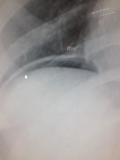
I suspected a PGU (perforated gastric ulcer), while the MO thought of maybe something else. Surgical referral was made, with chest + abdominal radiographs done. Chest X-Ray initially showed air under the diaphragm, while abdominal X-Ray showed suspicion of dilated large + small bowel. However, a repeated chest X-Ray with air syringed into the stomach via NG tube showed that the "air" became smaller, and thus surgical team concluded that it was unlikely. By this time, UFEME results returned, revealing blood, WBC's and nitrite. A provisional of UTI was made by the surgical team with referral to the medical team, but I thought still of PGU, my MO of perforated small bowel, another of PGU like me, while boss thought of the possibility of ectopic, though unlikely especially with negative UPT. She tried to get a radiological opinion of the initial chest radiograph, but no radiologists could be contacted at the time. And thus even though her clinical state has improved, her definitive "diagnosis" was still uncertain. Heck, even amylase was raised (almost to 250).

Question: Is it possible to have a "false positive" air under diaphragm? Googling failed me. Here's the initial radiograph:


3) Hm, another young male with no known history of medical illnesses came with tingling of fingertips and toes. Appeared hyperventilating. Tingling disappeared with reassurance to breath normally, just waiting for normal blood electrolytes investigation for discharge. Are today's men becoming baby boys? Hehe...This was the 2nd case of male hyperventilating from "anxiousness" in the week.

4) Elderly male came in with constitutional symptoms, and history suggestive of dysphagia, constipation & melena. But he was generally stable and actually quite cheerful. Examination was unremarkable, with no melena as well. Chest radiograph showed local consolidation in the middle zone of the right lung, and reporting was requested for expert opinion. How was the best way to manage the follow-up: specialist clinic/KK? The MO in charge opted for neither, telling him to come again to the green zone (!) in a week. This was to make it easier (and certainly faster than going to the KK) to present the radiological report from the radiologist and correlate it clinically.

Wednesday, May 27, 2009

ED Hell Continues

Today I was again at red, where I feel that I'm more and more like a Jonah (is that how we spell it?) in that zone.

ED Serdang is feeling more and more like KK Jaya Gading: med students have power to manage patients, the problem being that they're life and death patients and the doctors in-charge sometimes go MIA, forcing decisions to be made by med students and support staff. How about "Hello Mr. X, I'm your medical student in charge for today, literally" as the greeting of the day (of course I didn't say that)? Not being able to key in requests on behalf of missing doctors made things worse. And it's all because of a doctor seeming to have what I think personal problems, taking time to cry and weep for an unknown reason, before disappearing off someplace.

And the system of waiting for investigations before departments accept patients made things pretty stressful. There was one patient who had an obvious case of ruptured/impeding rupture of an ectopic pregnancy: generalized lower abdominal pain referred to the shoulder, difficulty in taking deep breaths, LMP with positive UPT and PV spotting with pallor. GP trans-abdominal scan showed no POC in-utero. But we had to wait for blood results which only returned 2 hours later: Hb was dropping down to 7. Then did the O&G MO see her, and found an adnexal mass with free peritoneal fluid. Of course she was sent to OT stat. I felt tempted to perform a cervical excitation test to just get her referred already.

How about over-triaging for political reasons? There was an elderly lady with 3 days of acute gastroenteritis, and clinically only mild dehydration and hyperventilation without Kussmaul breathing. Past history suggestive of BPPV without seeking medication, and HPT/DM/LVH on meds. The dagger? She was tolerating orally! She was here in red because she's the mom of the previous director. Only managed with maintenance fluids and nourishing fluids brought in by the ex-director (contradictory, no?).

Of course boss had to attend to her (being in red) with me accidentally stuck there as I attended her before the MO; better manpower could've been sent to the patient with CKD, decompensating CCF, DIVC with underlying septic shock. He was there from morning to evening, as departments argued where to send him, until ICU decided to accept him, but pending on waiting for a patient to be TO'd.

Had an interesting case of hypokalemic paralysis, but couldn't clerk her as I was busy seeing other patients and she was sent to the wards when I was a bit free.

Having to pick up the slack for the MIA doctor, Nu'aim and I only returned home at 6 pm, one hour late. Totally screwed my plan to go to the gym tonight, as I was completely drained, forced to "lead"* the management of a patient with hypoglycemic coma with almost impossible vascular access (got an ankle access on our 7th try), as there were no doctors around when he came around 5 pm. Soon after, the yellow zone doctors had to cover the red, while one of the green doctors shifted to yellow when they knew of the MIA doctor.

I felt some pity going back, but hey, you shouldn't overwork if you're on office hours duty instead of shifts.

But stress leads to good timely humor. Quote of the day of an ENT MA to an uncooperative patient a week back:

"Aiyoo, uncle, tolong duduk diam...Sudah pukul 5 lah! Saya mau balik..." (Uncle, please sit still, it's 5 already! I wanna go home!)

Or at least something like that. But seeing others post pics make me jealous, as I don't have a camera, and can't take pics anyway as there's no time to slack off from "work"; at least any sort of humor relieves the stress.

*Not a good thing, especially when other doctors/patients/relatives think that you're a doctor. There can't be responsibility without power, just as it goes the other way around.

Thursday, May 21, 2009

When Worrying Too Much is a Problem, Literally.

Interesting cases today (as thinking exercises for to-be final year students):

1. Middle-aged woman presented with clear mucus coughing + SOB, LOA, LOW for 2 weeks 3 days, associated with pleuritic chest pain, night sweats and shivers. On observation, her eyes are opened extremely wide, face is pale but pink conjunctiva, very anxious, a bit tachypneic (20-22 breaths per minute). But the best part is that despite dyspnea, her SpO2 was 100% under room air. While clerking further she complained of cramping sensation of the fingers. This is her second visit to ED. Spot diagnosis?

2. Middle-aged man with no known history of medical illnesses presented with sudden severe headache, giddiness, nausea and vomiting since 30 minutes. No SOB or chest pain. Lungs clear. ECG and capillary sugar were normal. Similar episode a few months ago, resolved on its own. Differential?

3. Ever seen the pain of a patient with classical renal colic? Writhing about moaning in pain, hand at loin. What should you do first after allergy screen?

4. Elderly woman with a history of hypertension for about 10 years presented with fever and cough for 2 days, associated with shortness of breath, pleuritic chest pain and disorientation. Examination revealed edema up to both knees, coarse crepitations in middle to lower zones bilaterally, with displaced apex beat. Provisional?

5. 2 month old baby boy presented with history of 1 week on and off fever and cough, rapid breathing, difficulty in feeding. Examination revealed tachypnea, subcostal retractions with rhonchi and crepitations. Impression?

6. 7 year old boy complained of left testicular pain. We didn't manage to see the patient, but what is the no 1 diagnosis to be assumed until proven otherwise?

7. Classical description of a dengue macular rash?


1. Hyperventilation with underlying pneumonia, probably CAP. The patient was so anxious about her pneumonia that she developed respiratory alkalosis during clerking, instantly relieved by breathing into a bag. Radiograph showed perihilar and lower zone haziness. Sputum for AFB negative.

2. Rule out cardiac causes. Always check the vital signs. This patient had hypertensive emergency (encephalopathy) revealed by taking the BP (I purposely did not reveal it the above even though it was obvious when checking the BP). Lowered the BP with oral nifedipine down to 140 systolic and he was completely back to normal.

3. Just give her analgesia and she will "shut up" (a good thing). From 10 to 3 pain score instantly.

4. CAP, to rule out decompensated CCF.

5. Bronchiolitis probably complicated with secondary pneumonia.

6. Torsion torsion torsion. Remember the location of both testes relative to each other (which one is higher) and correlate with the presentation of patients. It's clinical embryology? What if the left is higher than the right?

7. White islands in a red sea. Don't forget how to perform a Hess's test and it's interpretation.

Tuesday, May 19, 2009

ED Hell

Today was a f*ck day in Serdang ED if anything can be described as such.

The morning was relatively normal for me who was tagging in the red zone, while the yellow zone (where Nu'aim was) was completely swamped with unnecessary admissions, which I'll refer to later in another post. But all that changed when I returned for the afternoon.

First, we had some new patients that came during lunch break. But the morning MO didn't pass over the cases to the new MO, so we had to orient ourselves with the old cases on our own. Boss told the afternoon MO to help out with green, while the morning was finishing the morning work. As the morning MO was supposed to still be around, we expected her to handle the red zone while the afternoon MO dealt with patients in the green zone. While doing that, we were suddenly thrown into the frying pan. Mind you, all of the bays in the red zone were totally full.

First, a brought in dead patient (BID) was brought into the red zone. MVA, young adult, open fracture grade II of lt femur, ENT bleed visible. While documenting that patient, another came in. Middle age with chest pain, previous history of AMI with balloon angioplasty done. The MO was busy helping with green. The morning MO went to observation bay (OB) and literally "disappeared", so I had to clerk the patient.

While clerking the patient, a patient was transferred out, and another promptly transferred in from ambulance call. Young adult, eyes dilated and fixed, asystole; I assumed that the patient was BID as well. He was an industrial worker, who suffered injury by a machine falling on him. The MA asked me what to do with the patient, and I told him never mind as he's dead already. But I did promptly told the morning MO, for which she told me to tell the afternoon MO.

The afternoon MO was shocked as there was no proper passover, so I briefly told her about the BID patient, for which she (wrongly) decided to resuscitate. We even intubated him. Of course expectedly the patient wasn't revived. But I noticed that bagging the patient became progressively harder (I was in charge of the airway). The doctor also said that the bagging sounds were faint at best when she auscultated. I took a few seconds remembering anaesthesiology lectures. That's when it occurred to me: Shit! This patient has pneumothorax! And shittier still, none of us (MO, medical students, MA & nurses) properly examined this patient (we were still disoriented with the current patients, so were unprepared for new patients)!

I stopped bagging, and palpated the chest. Oh fuck, it was like a leather bag filled with hyperbaric air. Percussed - hyperresonant like those hollow watermelons.

"Dr, I think this patient has pneumothorax la."

The doctor palpated and percussed, as did the others. We gave her a gray (large bore) needle which she used to puncture the right pleura. Gas followed by blood. Bilateral tension hemopneumothorax.

Then we also noticed that his crotch was seen pretty big. We opened it up, and noticed a huge scrotal swelling. Intraabdominal injury. Then it occurred to us that there was also flank bruising. During intubation we also noted a mandibular fracture. We stopped resuscitation. Polytrauma, and anything might've caused his death.

I then reported about the patient with chest pain, and of course the MO was surprised. While going to attend that patient, boss asked us about the BID patient, for which she then scolded (especially) me and the MO for trying to resusc. a BID patient. In the middle of assessing the chest pain patient, another came in. Elderly in coma, hypoglycemic but not revived with dextrose. Family members not around (yet). Argh.

We tried to intubate him so that we could do a CT scan. I tried first as the MO offerred, and I forgot to assess the airway first (neither did she). Only while attempting did I notice that he was Mallampati IV! With TMD less than 6 cm.

As soon as we finished securing that patient, we saw another patient already in! Turns out that she was transferred from yellow zone, but the MO didn't inform us first (as we're busy resuscitating the BID patient). Elderly with apparently lowish GCS (even though the yellow zone MO said that she was 14/15, but we didn't listen to a proper history from the MO as we're busy already; yellow was also overflowing by then). Ooh, we were all well pissed off by then, and the afternoon MO was literally calling the morning MO to settle and pass over the morning cases (which we still knew almost nothing about yet in the red zone).

While being pissed, turns out that another was already in as well! 34 years with IHD, in for chest pain. I attended him, while the MO attended the elderly lady. As soon as we finished with both, the MO decided to review the morning cases by herself. Turns out that a morning patient didn't even have his blood taken. And no review ECG (k/c/o IHD, came in with SOB). Damn. I took the blood and ECG's of patients that required them, while the MO tried to review the morning cases and report the new patients.

Then she asked me to review the patient transferred from yellow. We were told that she had 14/15 GCS. Huh? E1, V3, M5 is not 14/15! While she tried to review the history taken by the yellow zone, I finished up the bloodtaking and ECG's.

5.30 pm, and I decided to call it a day.

And the worst part of the afternoon was that when I initially came into the red zone, the boss was with the HOD of medical dept. Something was wrong. PPE was ready to be used. I asked one of the MA's, "Ada suspected case ke (of H1N1)?"

He nodded. The patient was in the quarantine room, and while we're busy doing our thing, the boss was attending that patient with full biohazard regalia.

Lessons:
1) Always properly pass over/refer your patients.
2) Always properly expose unconscious patients with incomplete history. You can have med students and nurses+MA's to handle resuscitation, while the Dr makes a quick examination.
3) If intubation is done properly but still something is amiss (regarding (B)reathing, never ever forget pneumothorax (stressed by anaesthesiologists time and time and time again, as well as ED MO Dr Jeth)
4) Finish your work so that the next shift doesn't have to play catch up for you.
5) Serdang ED needs HO's. Like now. You can't have medical students who can't give input into the hospital IT-based recording system being the main (medical) helpers of the MO's.